pleural effusion cat ultrasound
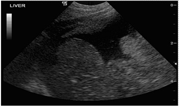
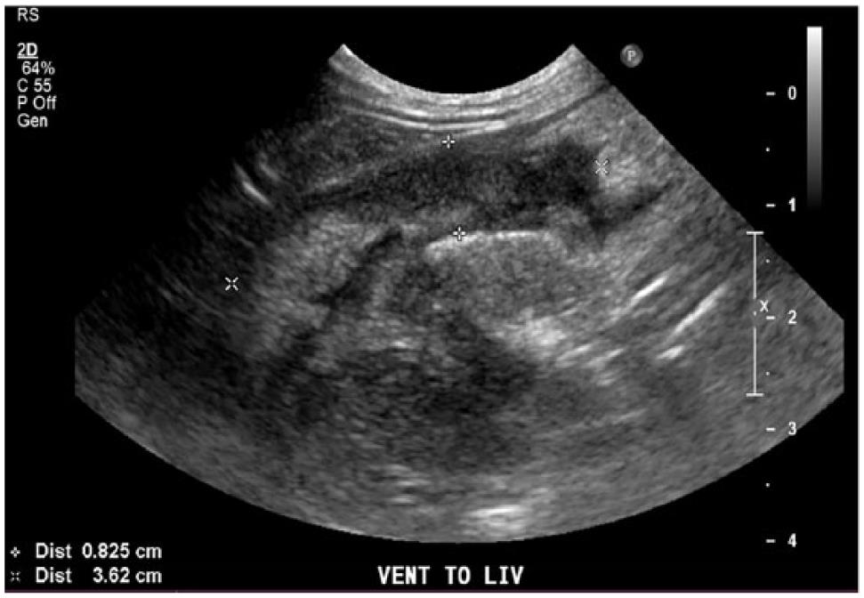
Pleural effusion is an abnormal accumulation of fluid within the pleural space and is a clinical manifestation of conditions such as pyothorax feline infectious peritonitis. Heres a labeled still image that still shows that spine sign.

Complete Guide To Feline Infectious Peritonitis Fip Clinician S Brief
Check out this video.

. Pleural effusion refers to the abnormal accumulation of fluid within the chest cavity. Dyspnea or respiratory distress. Reduced lung sounds due to fluid.
Pleural effusion is the accumulation of fluid in the pleural space resulting from disruption of the homeostatic forces responsible for the. Increased respiratory rate or tachypnea. In pleural effusion the fluid is not found within the lungs but instead within the pleural sac.
Four criteria have been described to differentiate ascites from pleural. Etiology Prevalence and Epidemiology. Point-of-care ultrasound is more sensitive than physical exam and chest.
Learn how to identify a pleural effusion with lung ultrasonographyDid you forget what normal lung looks like. Chest ultrasound has greatly improved the evaluation and interventional management of many pleural diseases. We review the literature on the use of point-of-care ultrasound to evaluate and manage pleural effusions.
This can be caused by thoracic lymphangiectasia swollen lymph vessels that leak chyle into the pleural. 1 Ultrasound has many advantages including the. Five cats without radiographic pleural effusion were later confirmed to have pleural effusion via thoracic ultrasound.
How the fluid came to be in the pleural space is tied in with this. Of the cats that received thoracic ultrasound most. A chest ultrasound to look.
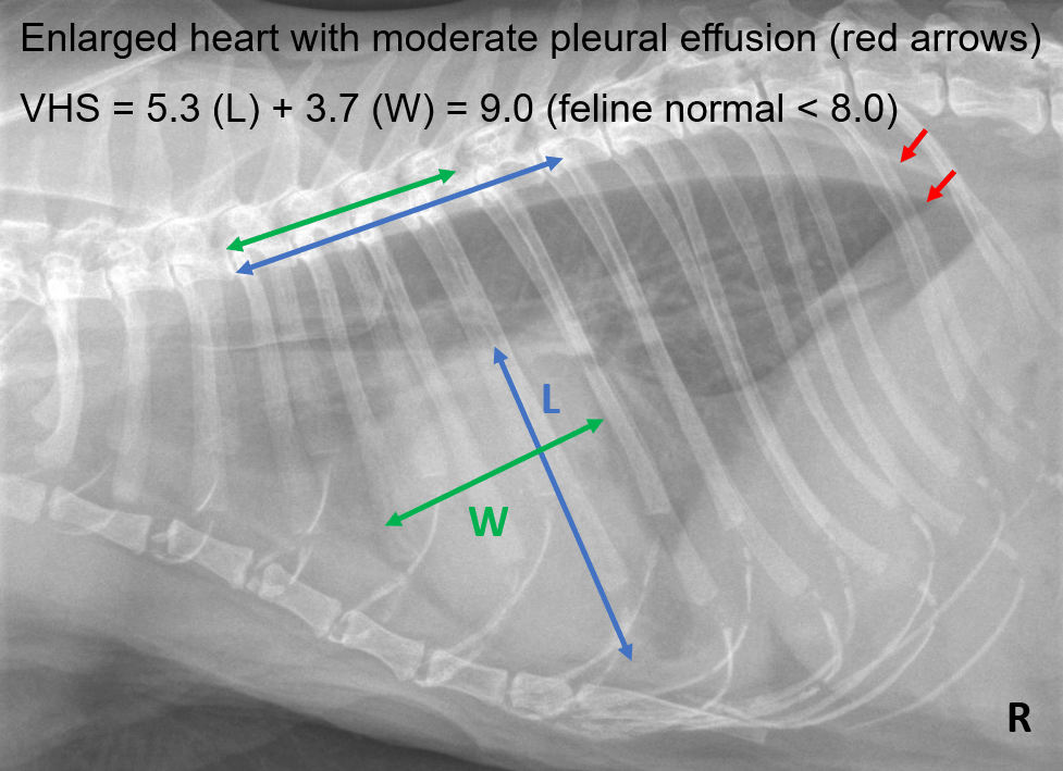
Both computed tomography CT and ultrasound US can be used to differentiate ascites from pleural effusion. The clinical signs of pleural effusion in the cat are generally as follows. There are a lot of causes of pleural effusion in cats transudate or exudate.
The most commonly diagnosed cause of pleural effusion in cats is chylothorax. Cats presenting with pleural effusion are nearly always in respiratory distress ranging from an increased respiratory rate and effort to open mouth breathing. Ultimately identifying a pleural effusion more quickly through handheld ultrasound examination rather than waiting for a chest X-ray may enable the delivery of faster patient care.
Its normal to see the spine behind the liver or the kidney but if you continue to see it up behind the thoracic space in back.

What Is Your Diagnosis In Journal Of The American Veterinary Medical Association Volume 258 Issue 2 2021

Severe Pleural Effusion In A Dog Affected By Larval Mesocestodiasis Sciencedirect
Focused Assessment With Sonography For Trauma Fast Mspca Angell

Search Imv Imaging
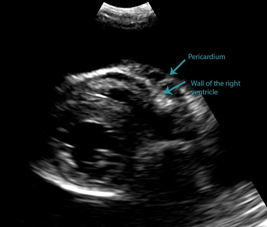
Differentiating Pericardial From Pleural Effusion Animal Ultrasound Association

Figure 1 From Caudal Mediastinal Thyroglossal Duct Cyst In A Cat Semantic Scholar
Case Study Hypertrophic Cardiomyopathy And Congestive Heart Failure In A Cat
Pleural Effusion In Cats Vetlexicon Felis From Vetlexicon Definitive Veterinary Intelligence

Frontiers Usefulness Of Chest Ultrasonography In Predicting Diagnosis In Non Emergency Small Animal Patients With Lung Parenchymal And Pleural Disease

Pdf Thoracic Ultrasound A Method For The Work Up In Dogs And Cats With Acute Dyspnea Semantic Scholar

How To Ultrasound Detection Of Pleural Fluid Case Study Video Youtube
Spontaneous Cholecystopleural Fistula Leading To Biliothorax And Sepsis In A Cat

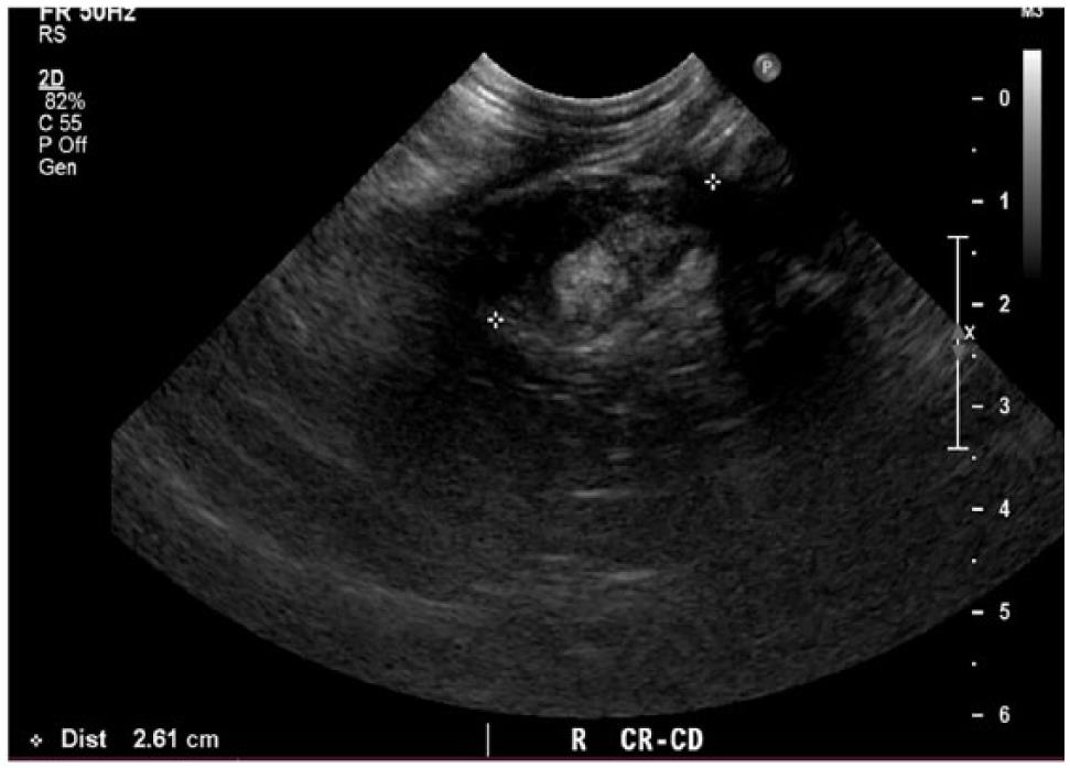
Different Types Of Pleural Effusion On Ultrasound Scan A Exudate B Download Scientific Diagram
Spontaneous Cholecystopleural Fistula Leading To Biliothorax And Sepsis In A Cat

Differentiating Pericardial From Pleural Effusion Animal Ultrasound Association

Fluid Colour Sign On Chest Ultrasonography In A Cat With Exudate Pleural Effusion And Pleuropneumonia Ho 2019 Journal Of Small Animal Practice Wiley Online Library
Pleural Effusion In Cats Vetlexicon Felis From Vetlexicon Definitive Veterinary Intelligence

Eosinophilic Pericardial Effusion In A Cat With Complex Systemic Disease And Associated Peripheral Eosinophilia Sciencedirect
![]()
Ultrasound Image Of A Large Thyroid Cyst In A Cat Note Cystic Fluid Download Scientific Diagram